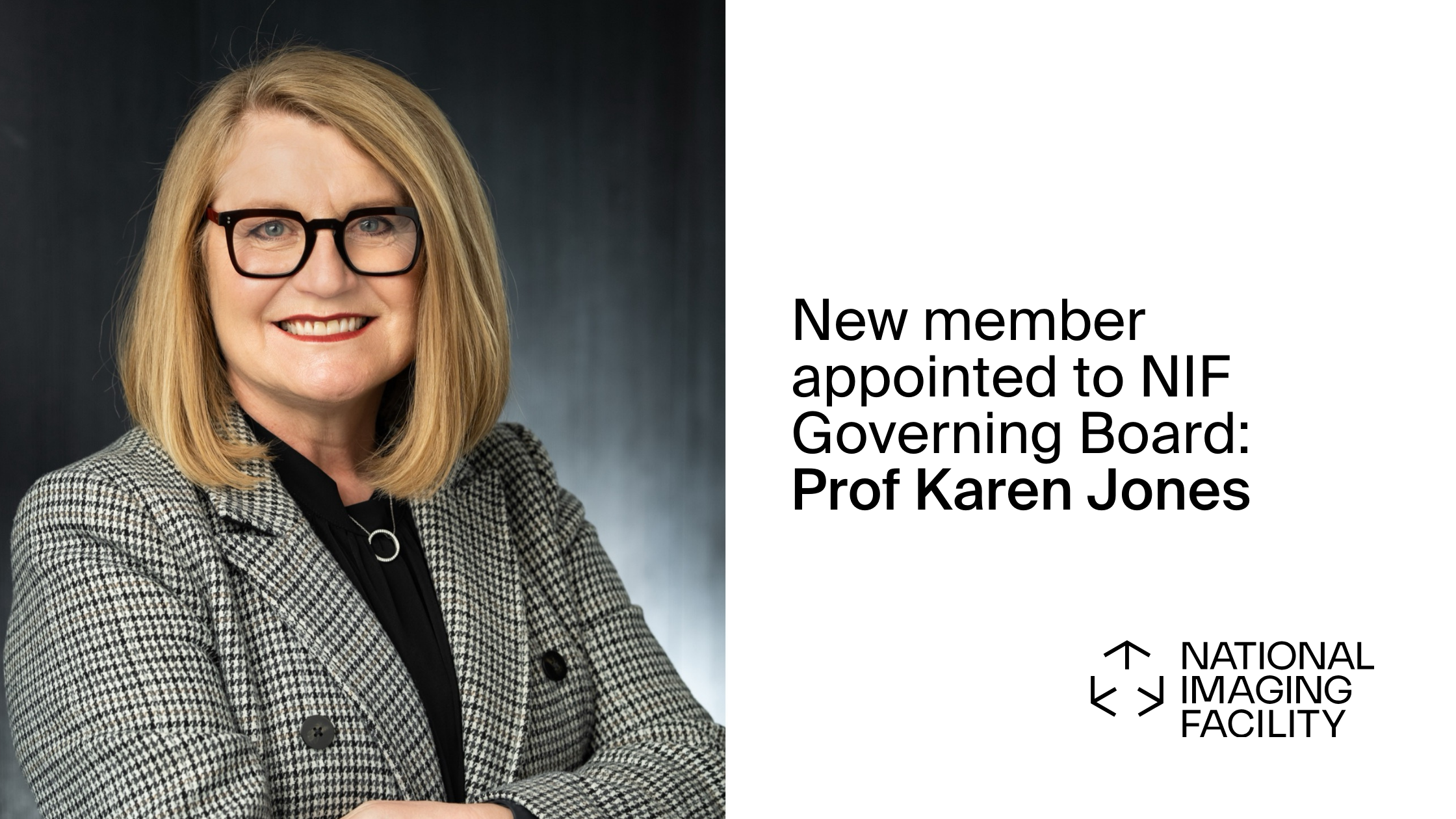
Announcement: Prof Karen Jones appointed to NIF Governing Board Posted on July 8, 2024July 28, 2024 by aburton
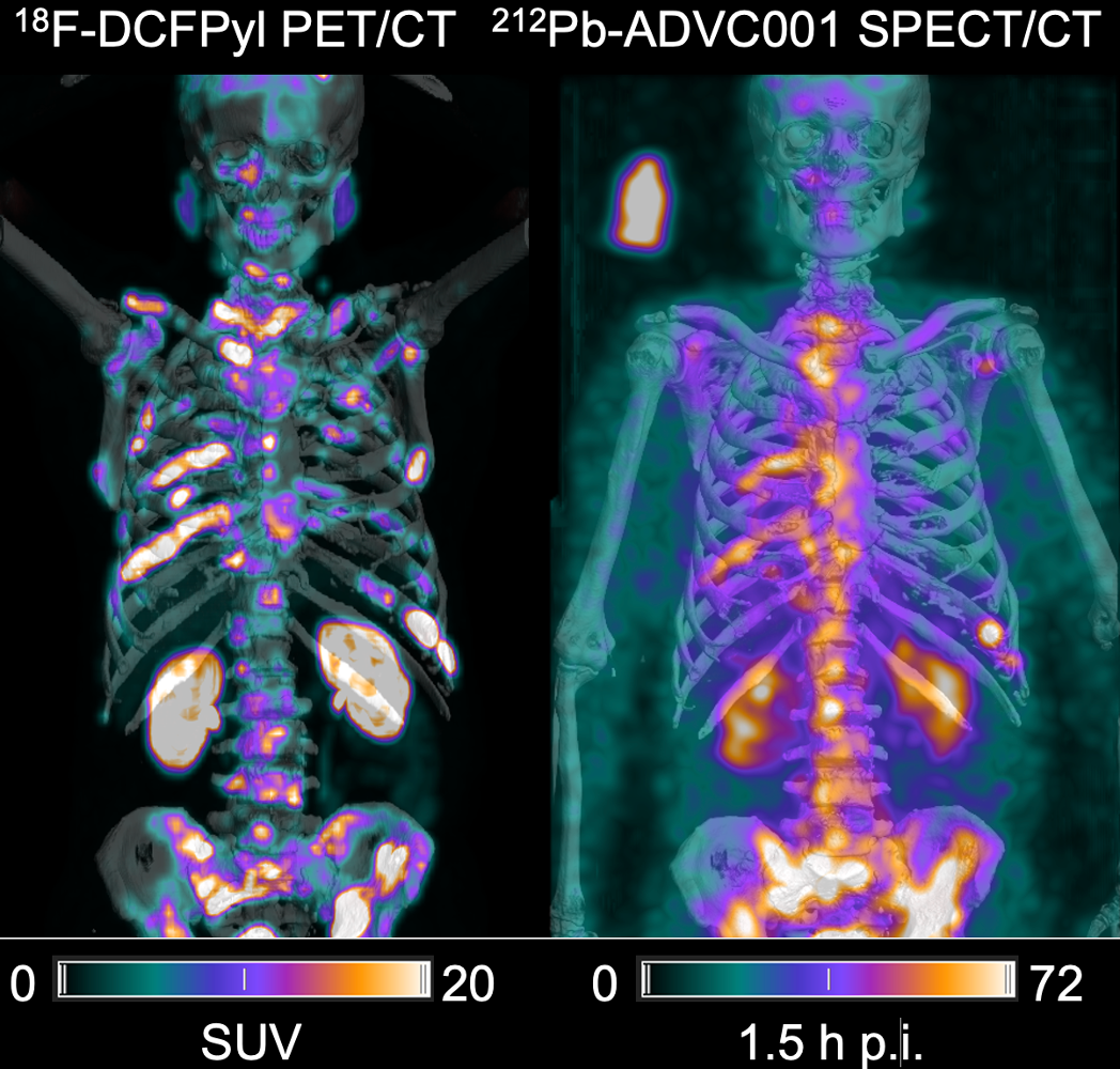
First in-human images of new hope treatment for poor-prognosis prostate cancer Posted on June 24, 2024December 17, 2024 by aburton
New brain imaging set to improve and speed epilepsy diagnosis, treatment Posted on June 24, 2024July 23, 2024 by aburton
National Imaging Facility receives $1.6m NSW Government funding to advance medical research Posted on June 24, 2024July 28, 2024 by aburton
Boosting health innovation and commercialisaton: $2m Queensland Government investment in National Imaging Facility Posted on May 14, 2024July 23, 2024 by aburton
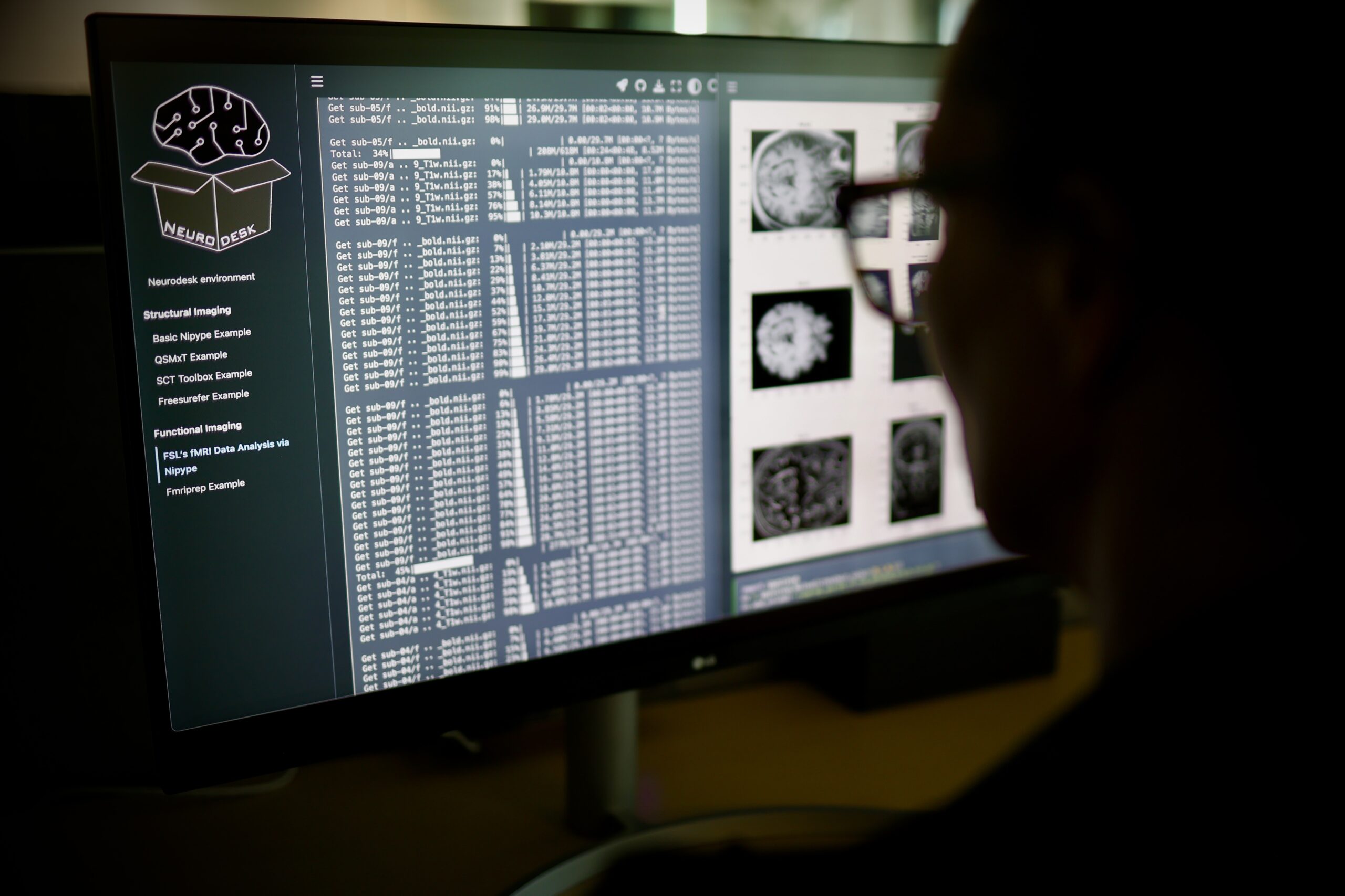
Brain research supercharged by new ‘control panel’ accessible worldwide Posted on February 6, 2024July 23, 2024 by aburton
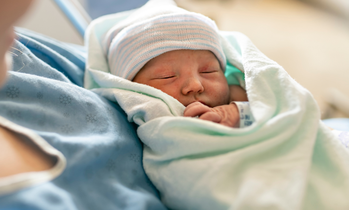
Revolutionary MRI technology unlocks solutions for foetal growth issues with minimal invasion Posted on December 18, 2023July 23, 2024 by aburton