The challenge
PET/CT imaging in pregnancy requires a delicate balance between diagnostic accuracy and minimising radiation exposure to both the patient and the foetus. Current dose-reduction strategies often compromise image quality, limiting their clinical usefulness. There is a clear need for improved imaging methods that significantly lower radiation burden while maintaining diagnostic accuracy.
Despite these concerns, vulnerable patients – such as pregnant women – may still require PET/CT scans for life-threatening conditions (such as cancer, infection, or inflammatory diseases) where precise imaging is essential for their diagnosis and treatment planning. There is a pressing need for imaging methods that reduce the radiation burden without sacrificing diagnostic integrity.
The solution
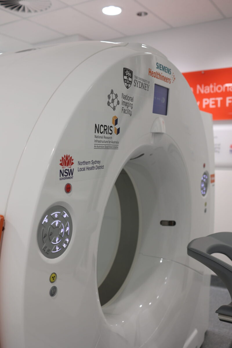
The Australian National Total Body PET Facility at the University of Sydney provides a unique platform that bridges clinical practice and research. It allows patients to access novel imaging strategies while accelerating their translation into real-world healthcare improvements.
By leveraging total body PET technology, the facility is exploring the feasibility of combining ultra-low dose PET scanning with cutting edge CT dose-reduction strategies, making PET/CT imaging more accessible for vulnerable patient groups.
As a result of a research-led initiative, a pregnant patient with lymphoma was scanned using an ultra-low dose FDG PET/CT protocol without compromising diagnostic usefulness. The high sensitivity of the total body PET system allowed for a five-fold reduction in radiotracer dose while maintaining PET image quality. Through a collaboration with Siemens, the study also used an ultra-low dose CT protocol, including the use of a tin X-ray filter, that enables accurate attenuation correction of the PET images and (further) reduced the patient’s radiation exposure from the CT scan by over 80%.
This work demonstrates how clinically embedding emerging imaging technologies can directly inform research and lead to rapid refinements in imaging protocols.
The impact
This application of total body PET offers an opportunity to validate ultra-low dose imaging protocols and contribute to future clinical guidelines for pregnancy. The findings may also improve radiation risk models and strengthen safety recommendations for medical imaging. Beyond pregnancy, the approach holds potential for paediatric patients and other dose-sensitive individuals, such as people who require frequent PET scans as part of long-term cancer treatment.
The increased sensitivity of total body PET further opens up new avenues for research in pharmacokinetics, first-in-human biodistribution studies, and radiobiology. This case highlights the broader role of total body PET in advancing imaging research and informing safer, more effective clinical practices.
“With the tremendous sensitivity and extended body coverage of this total body PET system, we get enormous flexibility in how we perform the vital scans that we provide. In this example, we were able to reduce the usual radiation dose to the pregnant patient by a factor of ten – to a level equivalent to the background radiation exposure received from living in Sydney for just 4 months. This was achieved without any compromise to the quality of the images, and the scan was obtained in 20 minutes.”
Clin Prof Paul Roach, Director, Department of Nuclear Medicine, Royal North Shore Hospital, Sydney