After 6 years and 762 scans, the world’s largest longitudinal study of children’s lower leg muscle volumes is complete. Dr Bart Bolsterlee and his team from Neuroscience Research Australia NeuRA – enabled by NIF – embarked on a study to answer fundamental questions about muscle growth and learn more about cerebral palsy.
The Muscle Growth in the Lower Extremity (MUGgLE) study examined muscle growth in typically-developing children and in children with cerebral palsy. Cerebral palsy is the leading cause of disability in childhood.
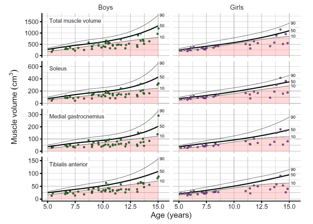
Using NIF facilities, 280 children, 80 of whom have cerebral palsy, had the muscles of their lower leg scanned up to 3 times over a period of 3 years to monitor their growth.
Reference curves – growth charts for individual muscles – have been created from the data, aiding the identification of children with underdeveloped muscles for their age.
Growth charts for muscles
The reference curves generated in the study can be thought of as growth charts for muscles, and used in a similar way as clinicians do for height and weight charts.
Symptoms of cerebral palsy are varied but often include muscle weakness and contractures – a stiffening of the muscle that limits joint range of motion. The reference curves in this study identified that 56% of the children with cerebral palsy had muscles that are small for their age.
“The targeted scanning and detailed image-analysis methods allowed us to isolate individual muscles – we found that 80% of children with cerebral palsy had at least one muscle which was small for their age,” Dr Bolsterlee explained.
“Now, we can say with confidence to what extent the muscles of an individual child are impaired. Almost all previous studies have only investigated muscle-group-level effects.”
Better informed treatments
The muscle-specific reference curves may become an important resource for clinicians and families to make informed decisions about treatment options for cerebral palsy and other conditions.
“The reference curves extract muscle-specific information, enabling a radiologist or orthopaedic surgeon to look at a scan and rapidly make more sense of it, aiding treatment choices. It could include the size of muscles, the architecture, the fibre orientations,” said Dr Bolsterlee.
“There are various treatment options for children with cerebral palsy but not all have been proven effective and some are controversial.”
“Limited data exists on how these treatments impact muscle growth in developing children, but I hope that the techniques that we’ve developed can start to change this.”
The research uncovered distinctions in muscle development between boys and girls. Initially, muscle growth overlaps until approximately age 11, after which girls experience a growth spurt, followed by boys two years later.
Additionally, the study revealed an asynchronicity between the timing of bone and muscle development, with bone growth peaking two years before muscle growth. These findings could be used to optimise the timing of medical treatments.
The importance of NIF’s MRI scanners and experts
Over 150 scans a year were carried out at NeuRA over the duration of the project.
“Having research-dedicated MRI infrastructure and support has been fundamental to our project,” said Dr Bolsterlee.
“It’s impossible to even think of doing a study of this scale at a clinical site – it would simply not be logistically feasible with the availability of scanners.”
“And having a dedicated research radiographer and help with optimising protocols – the knowledge is there and you take your research question and get expert help with the best possible way of answering that.”
Borrowing neurological techniques for muscle fibres
Muscles are composed of millions of muscle fibres, each the size of the human hair, typically several centimetres long and 10–100 microns in diameter. The challenge of visualising muscle fibres in the past is that they are too fine to be seen in an MRI scan.
However, Dr Bolsterlee and his team has been able to apply the technique of diffusion weighted imaging to muscle, which had been already extensively applied to analyse neuronal connectivity in the brain.
“By measuring the diffusion rates of water molecules in many different directions, you can reconstruct the orientation of the fibres in the muscles – it’s through this diffusion-weighted imaging that we can measure the fibre orientations in living human muscles in three-dimensional detail,” Dr Bolsterlee explained.
The MRI images were automatically segmented by using deep learning of convolutional neural networks – to human-level accuracy, in a matter of minutes.
Until recently, quantifying muscle volume from MRI images involved manual segmentation of muscles – the time taken to do this on hundreds of scans to create reference curves would have been nearly impossible without AI.
Given that skeletal muscle constitutes approximately 40% of adult body weight, understanding our muscles has far-reaching significance – for diseases, development, and sport science.
And for children with cerebral palsy and other conditions affecting muscle growth, adding to the body of knowledge of the growth and mechanics of individual muscles will advance treatment options.
The journal paper about this research was published in PNAS and in the Journal of Anatomy.