#WomenInScience: A conversation with Diana Patalwala, Research Officer, Preclinical and Materials Imaging and National Imaging Facility Fellow at the Centre for Microscopy Characterisation and Analysis, University of Western Australia
11 February is the United Nations’ International Day of Women and Girls in Science, highlighting the importance of full and equal access and participation.
We’re proud to create powerful collaborations across the research and innovation sector, building teams with world-class expertise, who manage our state-of-the-art equipment, and partner with experts in other fields.
Our mission is to make cutting-edge imaging capabilities accessible to Australian researchers, and we envision a society that provides equal opportunity for people of all genders to learn, work and engage in science.
As we look to the future of research, it’s clear Australia’s success depends on us developing and encouraging the next generation of scientists, problem solvers and leaders – regardless of their gender, background or any other factor.
Today we highlight the exceptional work of women leading the way in science and thank them for their work to deliver the impacts of life-changing research.
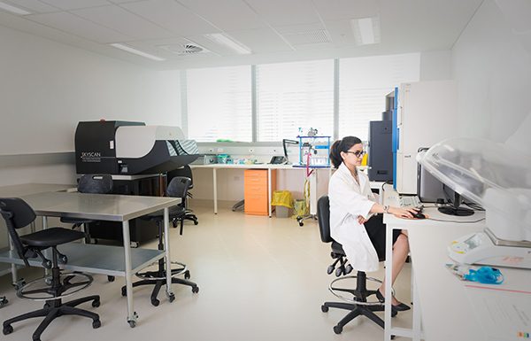
Diana Patalwala has worked with the National Imaging Facility (NIF) as a Facility Fellow at the University of Western Australia (UWA) for the best part of a decade, dedicating her time to enabling research translation to real-world benefits.
The breadth of impact that advanced imaging techniques has on research outcomes is what drives her to come to work every day.
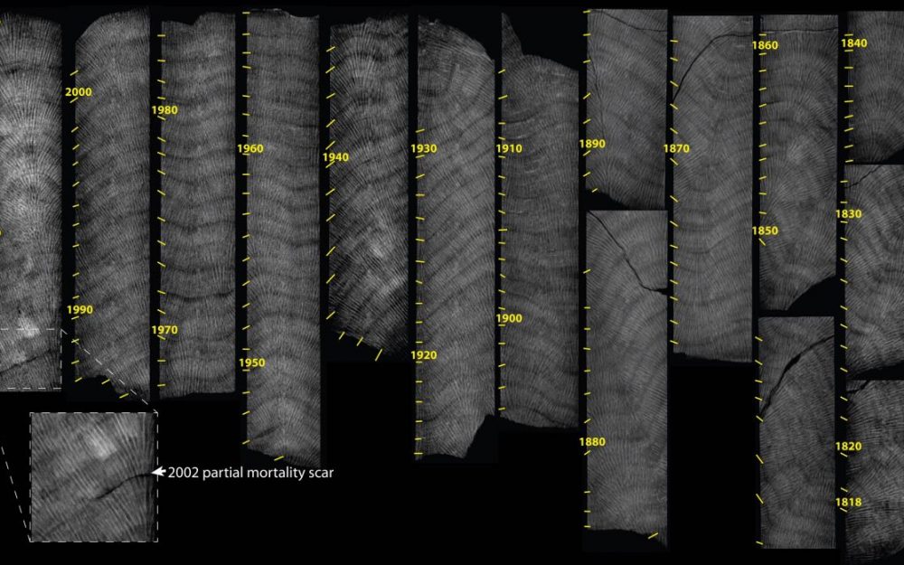
“We have researchers working on projects spanning in scope from investigating the anti-tumour effects of honeybee venom to treat breast cancer, the most common cancer in women worldwide, all the way to studying the acclimatisation of reef-building corals to consecutive heatwaves, contributing to the understanding of how different coral species are responding to climate change,” she says.
“This sort of research is contributing to society, it’s giving back, it’s impactful!”
Diana oversees the operations and development of research projects, providing user training and support at the Centre for Microscopy Characterisation and Analysis (CMCA) Bio-Imaging Facility (BIF), which supports interdisciplinary and multimodal imaging of small animals and materials using X-ray CT, High Frequency Ultrasounds and Photoacoustic Imaging, Fluorescence Multispectral and Bioluminescence Imaging.
Her valuable skills and experience in imaging methodologies enable her to assist researchers with data collection, reconstruction, analysis and visualisation.
When asked what led her to this career path, Diana says her post-graduate studies piqued her interest – but not in the way you might expect.
“My postgrad degree in Medical Biotechnology had a few units which involved data analysis from preclinical imaging instruments,” she explains.
“Although we were taught the theoretical principles on which these pre-clinical instruments worked, we were never allowed to operate them ourselves, which was disappointing because the science behind the instruments was really fascinating to me!”
“Seeing my professors at the university working with these instruments motivated me to envision my career in a pre-clinical imaging facility,” she says.
Now, Diana’s work allows her to have a hands-on role in imaging, enabling potentially life-changing research in medical biotechnology.
Before new medical treatments and drugs reach the clinical trial phase (when research studies are performed on people for evaluation), they undergo pre-clinical testing and development.
Diana says this is where pre-clinical imaging comes into the picture to provide invaluable data.
“High resolution and high throughput pre-clinical imaging equipment such as pre-clinical CT scanners, high frequency ultrasounds, photoacoustics, Invivo bioluminescence and fluorescence imaging techniques better facilitate the development of these treatments and drugs during their pre-clinical phase,” she says.
“As a NIF Facility Fellow, I operate and train researchers to use these instruments in a way in which we can get the maximum output from them and analyse the data they generate.”
Talking to Diana, it is clear she is extremely passionate about her job and how her work can benefit the research community.
At the end of last year, she presented her work on In vivo MicroCT and In vivo Fluorescence Imaging to an international audience at NIF’s webinar series in partnership with Global BioImaging, which for most people would be a career highlight – but for Diana, it’s quite a competitive ranking.
“EVERYDAY is a career highlight!” she says.
“Every day, researchers come to us with questions that have never been answered before, and we at NIF help them design experiments that give them access to world-class, cutting-edge pre-clinical and clinical imaging technologies.”
“We provide them a better insight into their research needs, and ultimately aim to generate answers to some of the biggest challenges facing society!”
When asked what advice she would give to someone who is considering working with a NIF capability, Diana says collaboration is at the heart of her work.
“Come and have a chat with us – we are here for you!”
“No one knows our instruments better than us – so talk to us before you design your experiments. We can put these instruments to use in ways you might not have thought of, and we will help you get the maximum output from them.” she says.