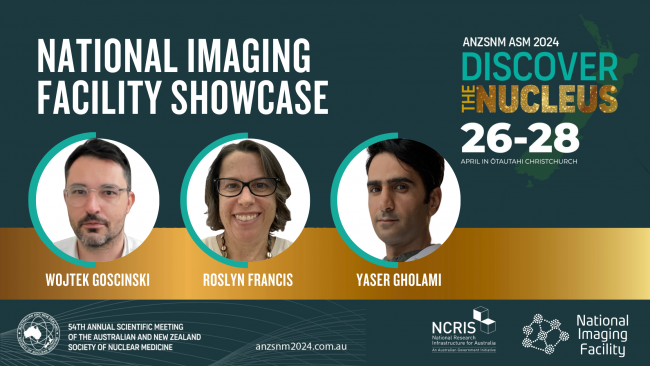
National Imaging Facility to host showcase session at ANZSNM 2024
National Imaging Facility will host a showcase session at the 2024 ANZSNM Annual Scientific Meeting, ‘Discover the Nucleus’ which will run from 26-28 April 2024 in Ōtautahi Christchurch.
Session details:
National Imaging Facility Showcase
Sunday 28 April
1:15pm – 2:00pm
Te Pae Auditorium
Speakers:
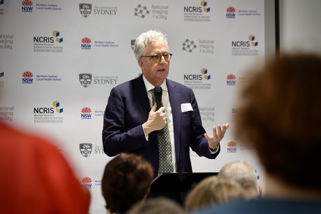
Prof Wojtek Goscinski
Introduction to National Imaging Facility
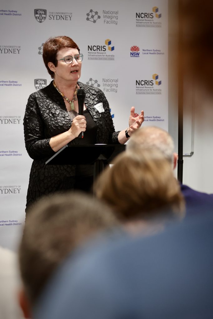
Prof Roslyn Francis
Toward an Australian Human Molecular Imaging Network
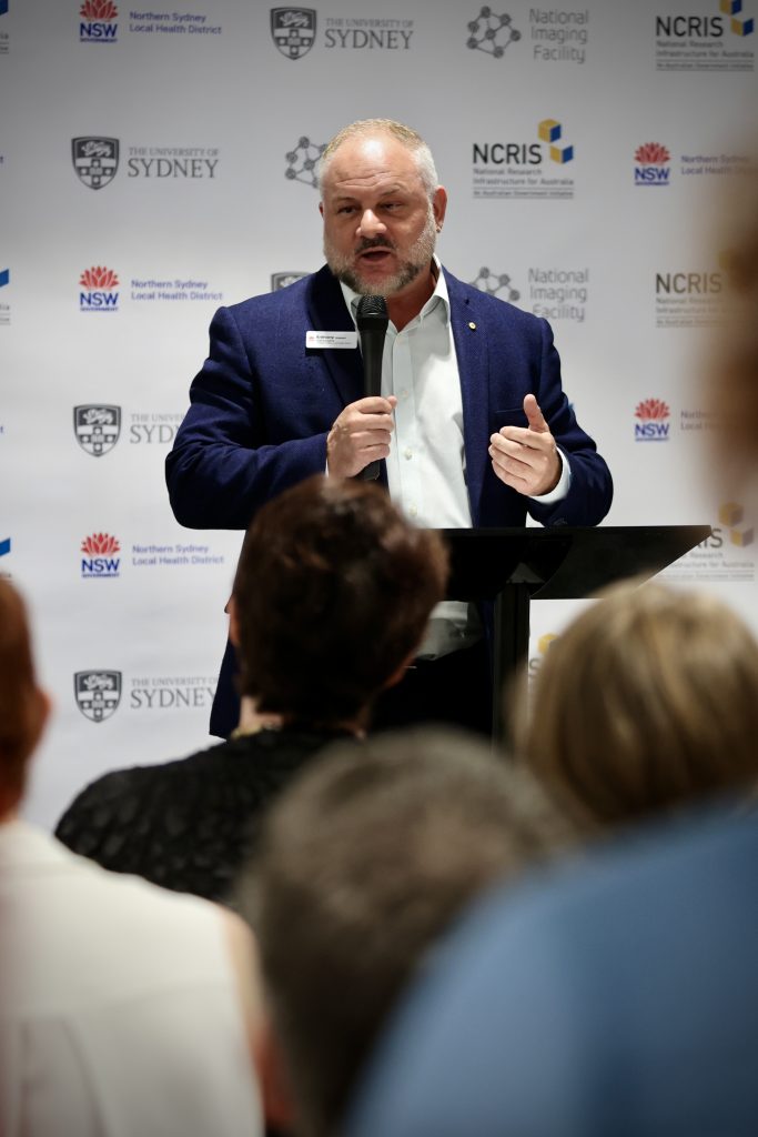
Dr Yaser Gholami
The National Imaging Facility PET Fellow Network
Click here for more information about the 2024 ANZSNM Annual Scientific Meeting and registration.