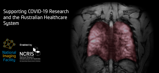
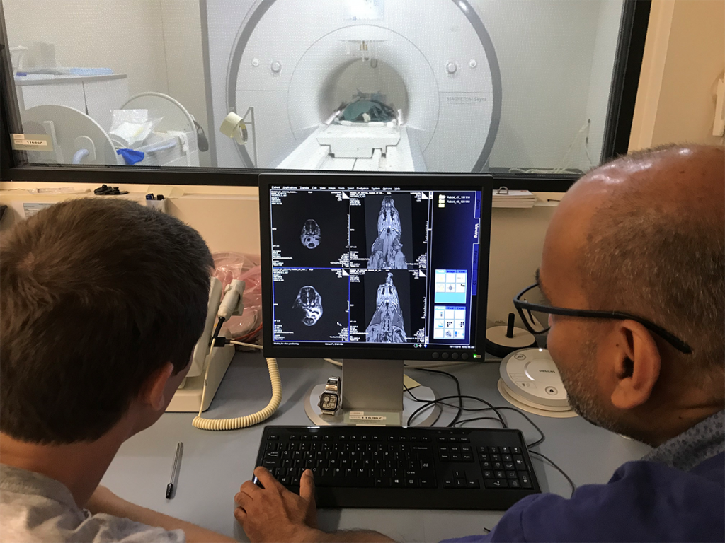
COVID-19 Research at LARIF: Using fluoroscopy for lung ventilation analysis

LARIF has teamed up with Australian biomedical company, 4DMedical, and University of Adelaide scientists Associate Professor David Parsons and Dr Martin Donnelly to address the COVID-19 crisis, through testing a novel ventilator, the now patented 4DMedical ‘XV technology’, and a large animal model of Acute Respiratory Distress Syndrome (ARDS).

Associate Professor David Parsons and Dr Martin Donnelly in the LARIF Cath Lab

The NCRIS-enabled facilities and expertise at the Large Animal Research and Imaging Facility (LARIF) NIF Node, located in the South Australian Health and Medical Research Institute (SAHMRI), were utilised by a consortium of doctors, engineers, and medical researchers as part of the Australian Lung Health Initiative (ALHI).

Read More

CT as part of the Forensic Science SA toolkit

A post-mortem examination, or autopsy, is a forensic technique for learning about the conditions of a person’s health at the end of life. These are typically carried out as part of a coronial inquiry to establish the cause of death. A post-mortem may also provide information about undiagnosed medical problems of relevance to family members and the research community.

Read More

National Preclinical PET QA

The NIF Molecular Imaging & Radiochemistry (MIR) Theme is a group of NIF Fellows, Directors, and users of NIF facilities that focus on state-of-the-art radiochemistry and molecular imaging applications using PET, SPECT, and MRI.

Integrating preclinical PET systems into a national resource requires the development of defined QA programs to monitor and integrate the data from individual systems. Hence, the MIR Theme initiated a national quality assurance (QA) program for the NIF preclinical PET instruments.

Read More

Impact of surgical lymph node removal

The impact of surgical lymph node removal on metastatic disease and the response to immunotherapy

Surgical resection of cancer remains the frontline therapy for millions of cancer patients every year, but disease recurrence after surgery is common with a relapse rate of around 45% for lung cancer. Relapse rates are expected to decline, with new immunotherapies producing extraordinary successes in several solid cancers. Immunotherapy administered after surgery could potentially ‘mop up’ small persisting cancer deposits that lead to disease recurrences. However, uninvolved (tumour-free) draining lymph nodes are the primary ‘factory’ for generating anti-cancer T cell responses; hence, should they be removed, subsequent immunotherapy may be negatively impacted. The aim of this project is to determine in murine models if the response of metastatic disease to immunotherapy is reduced following tumour lymph node resection.

Dr Vanessa Fear of the School of Biomedical Sciences, at The University of Western Australia, is investigating if the response of metastatic disease to immunotherapy is reduced following tumour draining lymph node resection. To do this, the Tumour Immunology Group is using an AB1 Model of metastatic disease. Tumour progression is visualised using IVIS imaging and histology following resection to track the effectiveness of treatment regimes. Ultimately, the team will seek to determine the impact of lymph node removal at the time of tumour resection to subsequent immunotherapeutic outcomes.

Fig 1: IVIS imaging from AB1-HA tumour model. Mice received AB1-HA_LUC i.v. and lung tumour development monitored on the InVivo Imaging System (IVIS, Lumina II imager). At the imaged timepoints mice received intrapertioneal injections of luciferin (150µg/g) and tumour burden was measured on the IVIS in photons/sec (p/s). A, tumour progression day 14 to day 19.

The research project involves collaboration with the Centre for Microscopy, Characterisation and Analysis, the West Australian Node for the National Imaging Facility to image, visualise and characterise the development of lung metastatic disease using the IVIS Lumina II in vivo bioluminescence imager with the help of Living Image Software (Caliper Life Sciences).

Fig 2: IVIS imaging and histology from AB1-HA tumour model.Comparison of IVIS reading with lung H&E staining showing AB1-HA tumour from the same mouse. Tumour volume determined using FIJI software.

The team have completed preliminary studies determining a 55% metastatic disease onset after surgical resection of the primary tumour. Current investigations in tumour resection and lymph node resection indicate temporal changes in onset of metastatic disease compared to mice with intact lymph nodes.

Further investigations into the impact of lymph node resection on immunotherapy are underway. Future investigations will include other models including lung adenocarcinoma, melanoma, and breast cancer.

Collaborators

School of Medicine, the University of Western Australia

School of Biomedical Sciences, University of Western Australia

Centre for Microscopy, Characterisation and Analysis, the University of Western Australia

This story was contributed by the University of Western Australia. For more information, contact Dr Vanessa Fear or Diana Patalwala.

NIF at BLiSS*Adelaide

BLiSS is a one-day event aimed at catalysing cross-disciplinary collaborations between early- and mid-career researchers. On Oct 4th 2019, BLiSS*Adelaide was launched, featuring exceptional lectures, poster sessions, and stalls of service providers and Universities. With over 150 registrations representing more than six institutions, the Adelaide scientific community came out in force to find new scientific collaborations. SAHMRI participated with excellent representation by Dr Randall Grose (ACRF research fellow), Dr Susan Porter (Manager PIRL) and Dr Marianne Keller (NIF Facility Fellow). Many of the posters featured data from SAHMRI’s small animal imaging equipment, and many attendees showed interest in the imaging modalities available through the NIF network such as high-resolution small animal MRI. For more information on NIF capabilities, contact Dr Keller or NIF Central.

Dr Randall Grose (ACRF research fellow), Dr Susan Porter (Manager PIRL) and Dr Marianne Keller (NIF Facility Fellow) at BLiSS*Adelaide


Magnetic Lymphatic Mapping in Pigs

Working Towards a New Gold Standard for Cancer Care


The lymphatic system is one of the key mechanisms for metastatic spread, whereby cancer cells that have disseminated from a primary tumour are taken up into lymphatic vessels and transported to other locations in the body, beginning with the lymph nodes. Conventionally, surgeons map the lymphatic flow from a tumour site into the first draining nodes – known as sentinel lymph nodes – using a Tc99m radiocolloid. Drainage pathways of tracers may be imaged before surgery with gamma cameras (lymphoscintigraphy), and individual nodes detected during surgery with handheld gamma probes. These sentinel nodes are the ‘canary in the coal mine’ and can offer valuable information to help stage the disease and determine the most appropriate treatment options, so it is ideal for removing just these nodes while leaving uninvolved nodes intact.

While this technique is successfully and routinely applied to breast cancer and melanoma, accuracy is limited in cancers where the nodes are in close proximity to the primary tumour or other nodes, such as cancers of the head and neck. Without an accurate method to identify sentinel nodes in oral cancer, extensive dissection of all nodes in the neck region is routinely performed to ensure any potential draining nodes are harvested, yet approximately 75 % of patients undergoing neck dissection are exposed to the complications and morbidity of this invasive procedure without clinical benefit.

Dr Aidan Cousins and Dr Giri Krishnan have been working as part of a collaborative project between the University of South Australia and the University of Adelaide to demonstrate a novel, high-resolution technique for lymphatic mapping using magnetic nanoparticle-based tracers. Studies conducted at the South Australian Health and Medical Research Institute (SAHMRI) involve the injection of the magnetic tracer to the oral cavity of female Large White pigs, which are then scanned with a 3.0T Siemens MRI located at the large animal research and imaging facility (LARIF) NIF Node. Post-injection scans give Dr Cousins and Dr Krishnan detailed anatomical information of the drainage patterns of the tracer, which is then used to plan the surgery. During surgery, a handheld magnetometer probe developed by Dr Cousins is used, along with MRI data and visual identification, to pinpoint the draining nodes of interest from other, uninvolved nodes.

Two men looking at a screen in front of an MRI

Dr Cousins with Raj Perumal (LARIF) examining an MRI scan of a pig following injection of the magnetic tracer

The results of this experiment showed the magnetic alternative to be adept for mapping lymphatic drainage in complex environments. Triangulating the location draining nodes with high precision before surgery was made possible by the high-resolution soft-tissue detail afforded by the 3.0T MRI. During surgery, the handheld probe was able to identify all draining nodes by way of detecting their magnetic ‘signature’. This final confirmation of draining nodes is analogous to the use of handheld gamma probes but has the distinct advantage of pin-point resolution, meaning nodes can be in very close (touching) proximity to each other and it is still possible to differentiate between the individual nodes’ magnetic signals. This spatial resolution is currently unmatched by any other probe technology commercially available and is key to the application of sentinel node mapping in complex cancers.

Photo of the high-resolution magnetometer probe developed by Dr Cousins


Results from this study are being used to design a world-first clinical trial applying magnetic tracers and a high-resolution probe to human patients with cancer of the oral cavity.


This story was contributed by SAHMRI. For further inquiries, please contact Mr Raj Perumal

Radiographers visit LARIF

In conjunction with the Australian Society of Medical Imaging and Radiation Therapy (ASMIRT) conference, the International Association of Forensic Radiographers (IAFR) organised a site visit to the South Australian NIF Node, Large Animal Research and Imaging Facility (LARIF), on the 31st of March 2019 to view the facilities used for post-mortem imaging (CT & MRI). Presentations included the practical aspects of post-mortem imaging using CT & MRI by Mr Raj Perumal and using CT & MRI in forensic practice by A/Prof Neil Langlois.

The participants were keen to understand the CT & MRI protocols used by Forensic Science South Australia (FSSA). LARIF provides a unique opportunity for post-mortem MRI to assist forensic investigations and there was a lot of interest from the participants to learn about the technical aspects of forensic MRI imaging.

A group photo in front of NIF and IAFR banners

The day before, at the ASMIRT conference, presentations were given about imaging opportunities and translation research at SAHMRI including a site tour of the rodent imaging facility at SAHMRI North Terrace by Dr. Marianne Keller.


This story was contributed by LARIF. For more information, please contact Mr Raj Perumal

Privacy Settings
Youtube
Vimeo
Google Maps